
ETDRS Grid (OCT)
When reading retinal OCT, you can place an Early Treatment Diabetic Retinopathy Study (ETDRS) grid on the fundus image, anchored to a foveal center point you choose. The grid is projected onto every B-scan of the paired OCT volume. On each B-scan it marks the region to grade and the subfield each part of the scan belongs to, so annotations on the B-scans have an anatomical location.
Requirements
The grid is available automatically for any OCT volume (OPT) that the DICOM data registers to a fundus image (OP) in the same study. The registration comes from the per-frame Ophthalmic Frame Location Sequence (0022,0031), which also provides the millimeter scale of the fundus image. There is nothing to enable. On studies without this registration, the grid controls do not appear.
Projects that de-identify data at upload remove DICOM sequences, and this removes the registration. OCT volumes uploaded before the grid was released may also need reprocessing. Contact us if a study you expect to support the grid does not.
Placing the center point
Open the fundus series of an OCT study and click Place ETDRS center point at the bottom of the viewer. Then click the center of the fovea. Press Esc or click Cancel to stop without placing.
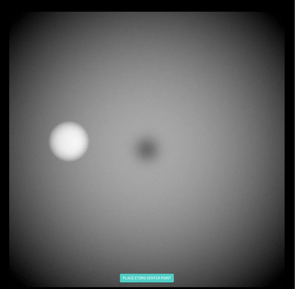
The grid is drawn around the point you clicked: a central subfield plus inner and outer rings, each divided into superior, nasal, inferior and temporal quadrants. To adjust the grid, drag the center point. The B-scans follow the grid as you drag, and the new position is saved when you release.
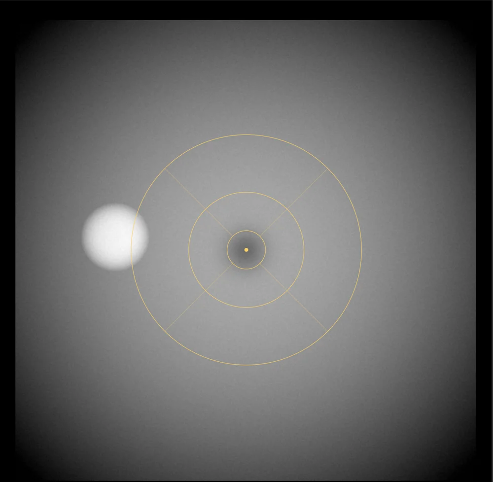
The center point is never placed automatically. Device estimates of the fovea are least reliable in the pathological eyes that most need grading, so the reader places it.
Subfields on the B-scans
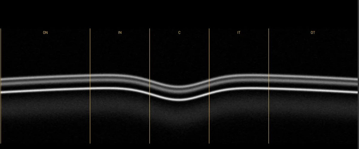
Each B-scan shows vertical boundaries where it crosses the grid, labeled with the subfield between them:
| Label | Subfield |
|---|---|
C | Central subfield |
IS, IN, II, IT | Inner ring: superior, nasal, inferior, temporal |
OS, ON, OI, OT | Outer ring: superior, nasal, inferior, temporal |
Here OS means outer superior, not oculus sinister. The eye is identified by the DICOM Image Laterality, which determines which side of the grid is nasal and which is temporal.
A B-scan through the fovea crosses all five horizontal subfields:
A B-scan above or below the fovea crosses only the rings it passes through. Here a superior B-scan crosses the inner superior subfield:
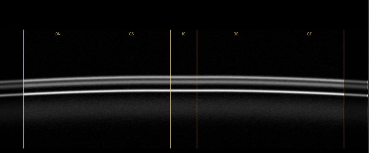
Near the edge of the grid, a B-scan may cross only the outer ring:
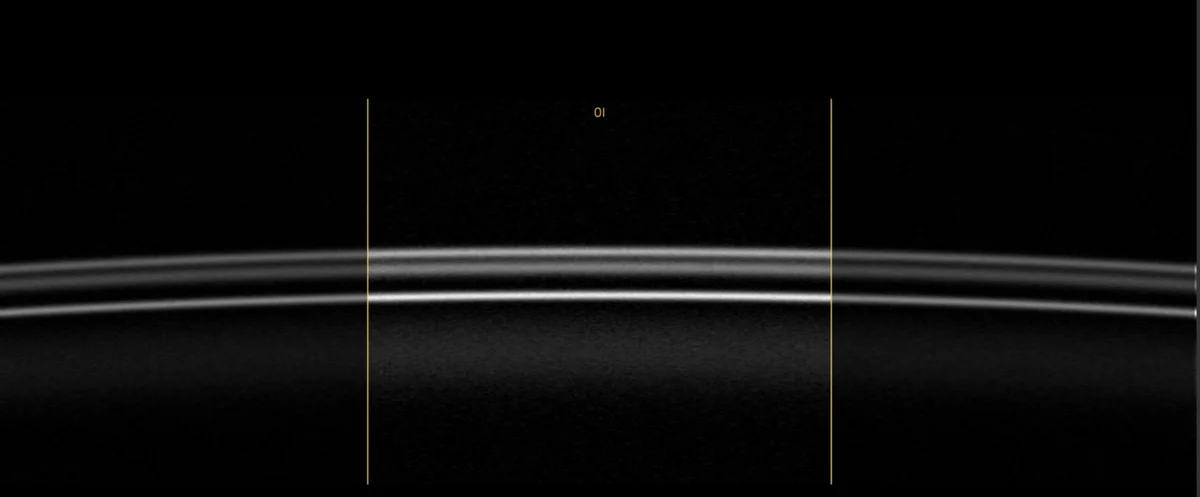
When a subfield is too narrow on screen to fit its label at the current zoom, the label is hidden. The boundaries are still drawn, so you can zoom in to read the label.
One grid per reader
Each reader places their own grid on each fundus image, and sees only their own. A QA reviewer adjusting their own grid does not move the grid of the reader whose work they are reviewing.
The grid is not an annotation. It does not use a label, and it does not appear in the annotation list. It defines the coordinate system that B-scan annotations are expressed in.
Showing and hiding the grid
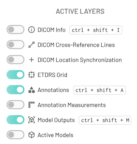
Toggle ETDRS Grid in the Layers control to show or hide the grid on the fundus image and B-scans. In a study with no registered OCT volume, the toggle is disabled, and its tooltip explains why.
Project settings
Project admins can hide the grid for the whole project or change the ring diameters. The default diameters are the standard 1, 3 and 6 mm. See Project Settings.
Export
Grids are included in the annotations export, as an ophthalmicGrids array on each dataset. Each grid is addressed by the DICOM UIDs of its fundus image, and records the millimeter scale it was placed with. You can reproduce subfield measurements from the export alone. See the JSON format.
Grids are not restored when annotations are imported into another project.