March 2022
Features
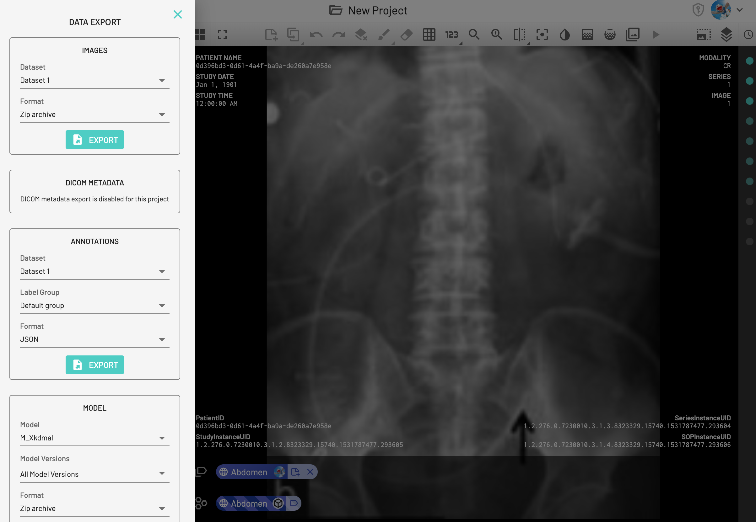
DICOM metadata export
DICOM files contain metadata that provides a huge amount of additional information about the image – for example image dimensions, modality used to create the data, and equipment settings. This metadata is organized into over 3,000 DICOM tags!
You may not always need this metadata, but when you do, MD.ai has introduced an easy-to-use DICOM metadata export function. Data is exported from the dataset(s) of your choice in a convenient normalized, human-readable JSON.
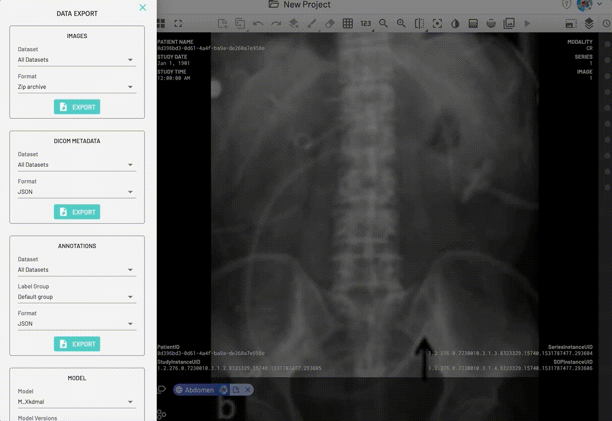
You can use your project’s metadata for a myriad of functions, for example keeping a large project on track by monitoring how many CXR images have been uploaded from each contributing site in a project, or using a script to track how many CTs are currently in a project.
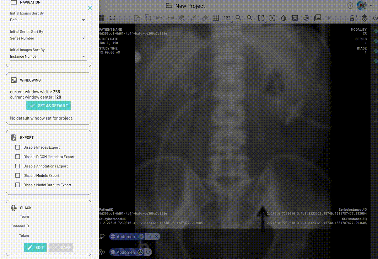
As always, with our convenient security and project management tools you can easily disable DICOM metadata export for your project if desired.
Allow duplicate UIDs on DICOM resource creation
DICOM files with identical UIDs but different content can now be loaded on MD.ai as long as they are loaded into different datasets! The DICOM standard, first published in 1993, allows effective interoperability between imaging systems. This extensive standard defines many rules including requirements for DICOM files to be uniquely specified by UID. While useful clinically, in testing and research settings users often desired to re-uploaded manipulated files without changing the UID which would cause image upload errors (for example image upload failed due to conflicting SeriesInstanceUID). This frequent concern has been resolved by scoping DICOM by dataset.
Bug Fixes
- Improved intermittent poor cine scrolling behavior by eliminating duplicate image loading calls, and by using explicit cine and fast series scrolling stop events to prevent stopping in the middle of scrolling.
- Fixed annotation import capabilities for multi-frame data – users can now upload annotation at frame level on multi-frame data (for example you can upload unique annotations on each frame of an Ultrasound video clip). For details on uploading via the API see our docs.
- Fixed copy annotation toolbar button functionality. Continue to easily copy annotations on one image of an exam to the next image (for example from image 4 to image 5 of your CT). Details on using the copy annotation functionality can be found here.
- Persistence of selected images when switching between datasets in Global Thumbnail Data View bug resolved.